
A segmentation method and system for intracranial aneurysm images

An intracranial aneurysm and image technology, applied in the field of medical imaging, can solve the problems of low segmentation accuracy and slow segmentation speed of intracranial aneurysm, and achieve the effect of improving segmentation accuracy and segmentation efficiency

Active Publication Date: 2019-03-08
UNION STRONG (BEIJING) TECH CO LTD
View PDF11 Cites 3 Cited by
  • Summary
  • Abstract
  • Description
  • Claims
  • Application Information

AI Technical Summary

Benefits of technology

This technology uses a method called central line based segmenting that automatically divides brain image data into smaller segments for easier analysis by computerized systems. It achieves these benefits through automated processing techniques with high precision and speed compared to existing methods such as manual sectioning or histology staining.

Problems solved by technology

This patents describes methods for improving the quality of medical imagery during diagnosis of brain damage due to strokes. However, current techniques are time-consuming and require skilled operators who have expertise over how well they work. There needs improvement in this process without sacrificing precision.

Method used


Image

  • A segmentation method and system for intracranial aneurysm images
  • A segmentation method and system for intracranial aneurysm images
  • A segmentation method and system for intracranial aneurysm images

Examples


Embodiment Construction

[0055]In order to enable those skilled in the art to better understand the technical solutions in this specification, the technical solutions in the embodiments of this specification will be clearly and completely described below in conjunction with the drawings in the embodiments of this specification. Obviously, the described The embodiments are only some of the embodiments of the present application, but not all of them. Based on the embodiments of this specification, all other embodiments obtained by persons of ordinary skill in the art without creative efforts shall fall within the scope of protection of this application.

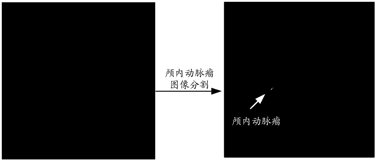
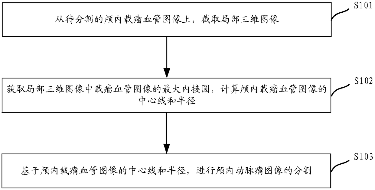
[0056] figure 1 A schematic diagram of a segmentation method for an intracranial aneurysm image provided for this specification. The method includes:

[0057] Step S101: intercepting a local three-dimensional image from the intracranial parent tumor image to be segmented.

[0058] DSA and MRA images are common images of intracranial aneurysms, but b...


PUM


Abstract

Embodiments of the present specification provide a segmentation method and system for intracranial aneurysm images. The method comprises the following steps of truncating a local three-dimensional image from an intracranial tumor-carrying vessel image to be segmented; obtaining a maximum inscribed circle of a tumor-carrying vessel image in the local three-dimensional image, and calculating a centerline and a radius of the tumor-carrying vessel image in the cranium; segmenting the intracranial aneurysm image based on the centerline and radius of the intracranial aneurysm-carrying vessel image.This method provides a segmentation method of intracranial aneurysm image with better segmentation precision and higher segmentation efficiency, and realizes the automatic segmentation of intracranialaneurysm image.

Description


Claims


Application Information

Patent Timeline
Owner UNION STRONG (BEIJING) TECH CO LTD
Learn More