An Osteomyelitis Lesion Analysis Method Based on Medical Image Registration

A medical image and analysis method technology, applied in the field of medical image processing and medical image registration analysis, can solve the problems of large differences in images, difficult to make judgments, inconsistent patient posture and occlusal relationship, etc.

Active Publication Date: 2022-01-28
SICHUAN UNIV
View PDF16 Cites 0 Cited by
  • Summary
  • Abstract
  • Description
  • Claims
  • Application Information

AI Technical Summary

Problems solved by technology

However, due to the inconsistency of the patient's posture and occlusal relationship when collecting data at different times, the images collected in different periods are very different
In addition, due to the different parameter settings such as window width and window level when the image is displayed, it is often difficult for the naked eye to distinguish the difference of subtle lesions, so it is difficult to make a very accurate judgment

Method used


Image

  • An Osteomyelitis Lesion Analysis Method Based on Medical Image Registration
  • An Osteomyelitis Lesion Analysis Method Based on Medical Image Registration
  • An Osteomyelitis Lesion Analysis Method Based on Medical Image Registration

Examples


Embodiment Construction

[0031] All features disclosed in this specification, or steps in all methods or processes disclosed, may be combined in any manner, except for mutually exclusive features and / or steps.

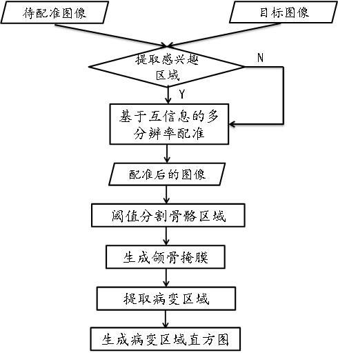
[0032] figure 1 Be the complete flowchart of the technology of the present invention, below in conjunction with Figure 2-Figure 7 The present invention is described in detail:

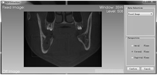
[0033] 1) Firstly, the multi-resolution registration method based on mutual information performs pairwise registration on the CBCT image data sets collected at different times before and after surgery; one of the data sets is taken as the original data set, and the other is used as the target data set; figure 2 It shows the two sets of CBCT image sets collected and the fusion display of the two sets of image sets before registration. From the left picture, it can be clearly seen that the jaw position of the two sets of images deviates;

[0034] In order to avoid the problem of inability to register or incomplete r...


PUM


Abstract

The invention discloses an osteomyelitis lesion analysis method based on medical image registration. The present invention extracts the lesion area of ​​osteomyelitis through accurate registration of images collected in different periods before and after operation, and allows doctors to intuitively and accurately observe and judge the development of the lesion at the same position through synchronous display browsing, gray value measurement and other technologies Recovery after surgery. The present invention includes the following steps: 1) Firstly, two-by-two registration is performed on the CBCT image data sets collected at different times before and after the operation using a multi-resolution registration method based on mutual information; one of the data sets is taken as the original data set, and the other is used as the target Data set; 2) In the registered image, the lesion area is extracted by sampling and analyzing the difference between the two images; 3) The lesion area is analyzed using the histogram and the evaluation result is output. The overall flowchart of the method is shown in Figure 1. The invention has the beneficial effect of greatly saving the diagnosis time of doctors, and has great application prospects in clinical diagnosis.

Description

technical field [0001] The invention relates to the technical fields of medical image processing and medical image registration analysis, and mainly relates to a method for analyzing osteomyelitis lesions based on medical image registration. Background technique [0002] In cranio-maxillofacial surgery, osteomyelitis is a refractory inflammatory disease commonly found in the upper and lower jaws, and it is often difficult to diagnose at the early stage of the disease. Usually, in order to evaluate changes in the condition, doctors will observe and compare CT / CBCT images collected in different periods, and make evaluation judgments based on their own experience. However, due to the inconsistency of the patient's posture and occlusal relationship when collecting data at different times, the images collected in different periods are very different. In addition, due to the different parameter settings such as window width and window level when the image is displayed, it is ofte...

Claims


Application Information

Patent Timeline
Patent Type & Authority Patents(China)
IPC IPC(8): G06T7/00G06T7/11G06T7/136G06T7/30
CPCG06T7/0012G06T7/11G06T7/136G06T7/30G06T2207/30008G06T2207/20104
Inventor 郭际香吕建成汤炜
Owner SICHUAN UNIV
Why Eureka
Try Eureka