Endoscope endcap for suturing tissue

a tissue and end cap technology, applied in the field of endoscopically suturing tissue openings, can solve the problems of unwanted and sometimes deadly infections, and achieve the effect of simple, reliable and controllable suture placemen

Active Publication Date: 2013-02-19
COOK MEDICAL TECH LLC
View PDF34 Cites 7 Cited by
  • Summary
  • Abstract
  • Description
  • Claims
  • Application Information

AI Technical Summary

Benefits of technology

[0006]The present invention provides medical devices, systems and methods for suturing a perforation in tissue, that may be used endoscopically and / or laparoscopically, and that offer simple, reliable and controllable placement of sutures around a perforation for complete closure thereof. One embodiment of a medical device, constructed in accordance with the teachings of the present invention, generally includes an endcap for use with an endoscope to suture an opening in tissue using a tissue anchor. The endcap has a tubular shape defined by an annular sidewall, and the sidewall defines an interior space. A side port is further defined by the annular sidewall and is in communication with the interior space. The side port is sized to receive and locate the tissue within the interior space for suturing. A support rib is positioned within the interior space and distal to the side port. The support rib extends laterally from a first position on the sidewall to a second position on the sidewall. The support rib and sidewall define a piercing aperture therebetween which is sized to be larger than a length of the tissue anchor, allowing the anchor to freely exit the interior space via the piercing aperture and side port.

Problems solved by technology

Openings or perforations in the walls of internal organs and vessels may be naturally occurring, or formed intentionally or unintentionally.
At the same time, there remain challenges to transluminal procedures, including providing a suitable conduit to the openings and body cavities, robust medical devices that are maneuverable via the conduit and operable within the body cavity, sterility of the conduit, maintaining insufflation of the body cavity, proper closure of the opening, and prevention of infection.
Travel of bacteria laden fluids outside of the gastrointestinal tract may cause unwanted and sometimes deadly infection.

Method used


Image

  • Endoscope endcap for suturing tissue
  • Endoscope endcap for suturing tissue
  • Endoscope endcap for suturing tissue

Examples


Embodiment Construction

[0019]In the present application, the term “proximal” refers to a direction that is generally towards a physician during a medical procedure, while the term “distal” refers to a direction that is generally towards a target site within a patient's anatomy during a medical procedure.

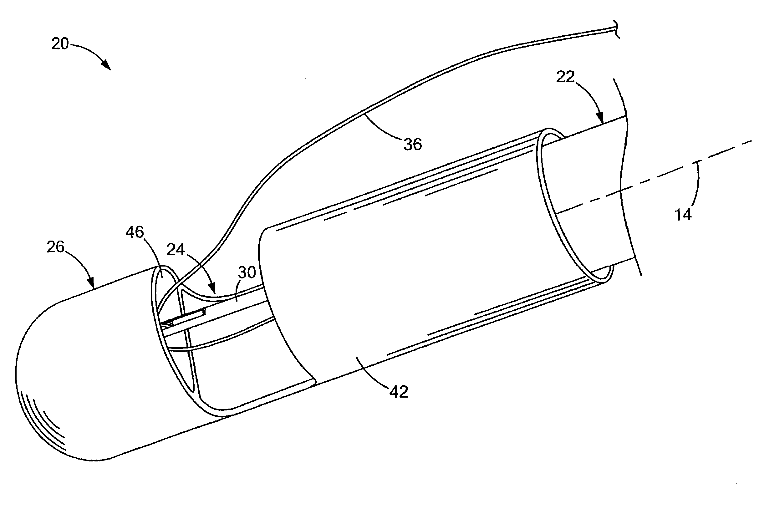
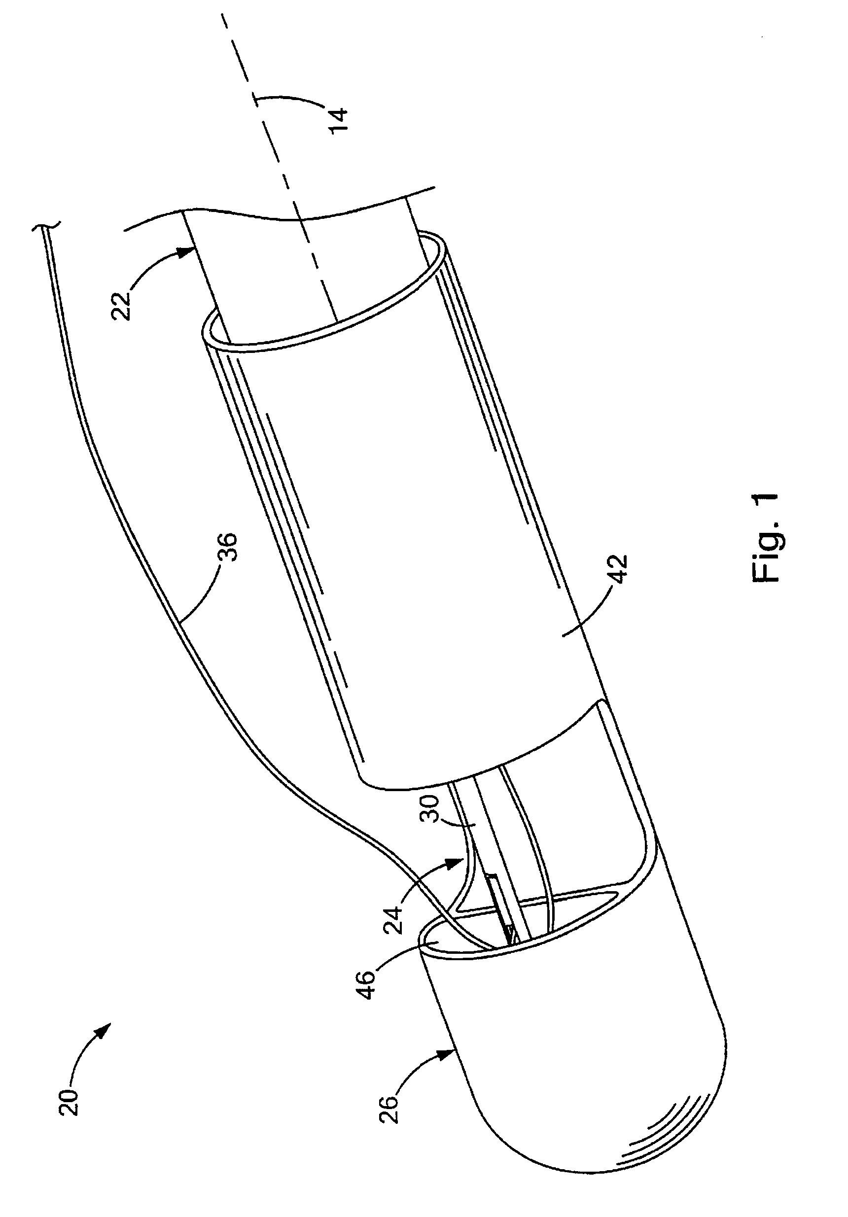
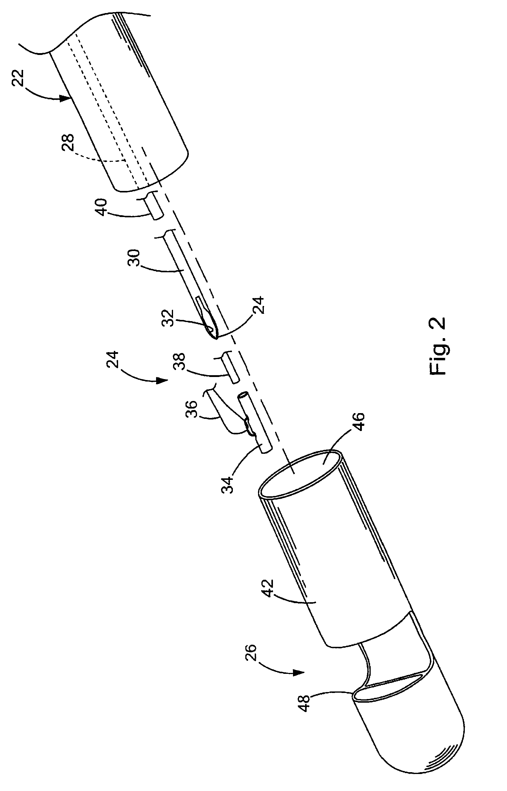
[0020]Turning now to the figures, FIGS. 1-2 depict a medical system 20 for suturing closed a perforation 10 in tissue 12 (see, e.g., FIG. 6), constructed in accordance with the teachings of the present invention. The medical system 20 generally comprises an endoscope 22, a needle assembly 24 and a medical device 26 adapted for use with the endoscope 22. The endoscope 22 may be any scope known to those skilled in the art, and therefore may have various lengths, diameters and functionality. The endoscope 22 generally defines a longitudinal axis 14, and a working channel 28 extends longitudinally through the endoscope 22. The needle assembly 24 is received within the working channel 28, and as best seen in FI...


PUM


Abstract

Medical systems, devices and methods are disclosed for suturing a perforation in tissue, that may be employed endoscopically and / or laparoscopically, and that offer simple, reliable and controllable placement of suture around a perforation for complete closure thereof. One embodiment of the medical device generally includes an endcap for use with an endoscope to suture an opening in tissue using a tissue anchor. The endcap has a tubular shape defined by an annular sidewall, and the sidewall defines an interior space. A side port is further defined by the annular sidewall and is in communication with the interior space. The side port is sized to receive and locate the tissue within the interior space for suturing. A support rib is positioned within the interior space and distal to the side port. The support rib and sidewall define a piercing aperture therebetween that supports the tissue being sutured.

Description

CROSS-REFERENCE TO RELATED APPLICATIONS[0001]This application claims the benefit of U.S. Provisional Application Ser. No. 61 / 109,337 filed on Oct. 29, 2008, entitled “ENDOSCOPE ENDCAP FOR SUTURING TISSUE” the entire contents of which are incorporated herein by reference.FIELD OF THE INVENTION[0002]The present invention relates generally to medical systems, devices and procedures for suturing tissue, and more particularly to endoscopically suturing openings in tissue.BACKGROUND OF THE INVENTION[0003]Openings or perforations in the walls of internal organs and vessels may be naturally occurring, or formed intentionally or unintentionally. These openings may be used to gain access to adjacent structures of the body, such techniques being commonly referred to as transluminal procedures. For example, culdoscopy was developed over 70 years ago, and involves transvaginally accessing the peritoneal cavity by forming an opening in the cul de sac. This access to the peritoneal cavity allows m...

Claims


Application Information

Patent Timeline
Patent Type & Authority Patents(United States)
IPC IPC(8): A61B1/00
CPCA61B1/0008A61B1/00087A61B17/0401A61B1/018A61B2017/0417A61B17/0487A61B2017/00296A61B2017/0409
Inventor HASHIBA, KIYOSHISURTI, VIHAR C.
Owner COOK MEDICAL TECH LLC
Why Eureka
Try Eureka