
Fusion protein and applications thereof

a technology of fusion protein and protein, applied in the field of immunotherapy, can solve the problems of serious adverse effects, cytokine storms, and even life-threatening reactions, and achieve the effects of less influence, excellent differential toxicities, and remarkable killing ability

Inactive Publication Date: 2019-11-28
CRAGE MEDICAL CO LTD
View PDF1 Cites 0 Cited by
  • Summary
  • Abstract
  • Description
  • Claims
  • Application Information

AI Technical Summary

Benefits of technology

The present invention provides an immune effector cell that can be effectively killed by a specific antibody-drug conjugate. This is achieved by expressing a fusion protein on the surface of the immune effector cell. The substance does not have substantial non-killing effects against cells that do not express the fusion protein.

Problems solved by technology

However, with the development of clinical trials of CAR-T cell therapy, there are many serious side effects, such as cytokine storms, off-target effects, etc.
When serious adverse reactions occur, if the CAR-T cells are not inhibited in time, serious adverse, even life-threatening reactions will be incurred.
However, since HSV-TK produces immunogenicity in patients, and patients receiving cell therapy will not be able to continue to use ganciclovir as an antiviral drug, both of which greatly limit the clinical use of HSV-TK. iCasp9 induces apoptosis of T cells expressing iCasp9 suicide gene by applying a small molecule drug (AP20187) in a patient.
However, AP20187 has not been commercialized, thus limiting the popularity of iCasp9 suicide gene.
When the complement system or NK cell activities in a patient's body is defective, killing effects of marker genes are often limited.

Method used


Image

  • Fusion protein and applications thereof
  • Fusion protein and applications thereof
  • Fusion protein and applications thereof

Examples


example 1

n of Fusion Protein FR806

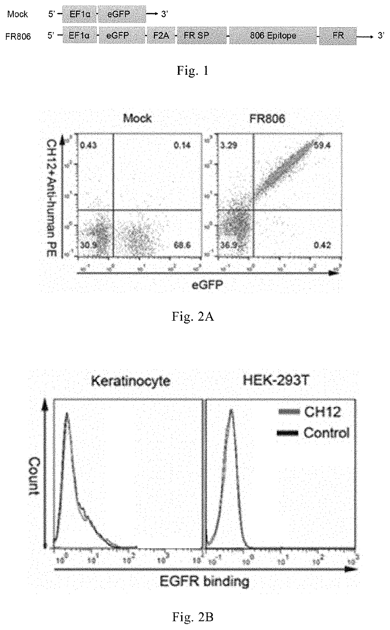
[0118]In this example, eGFP (enhanced green fluorescent protein) was selected as a fluorescent marker for analysis. F2A was selected as a self-cleaving sequence, and F2A is a core sequence derived from 2A of foot-and-mouth disease virus (or “self-cleaving polypeptide 2A”) and has a “self-cleaving” function of 2A; partial amino acid sequence (SEQ ID NO: 32) of human folate receptor of subtype 1 (FOLR1) and partial sequence of EGFR (SEQ ID NO: 28) were selected and expressed as a fusion protein FR806 (SEQ ID NO: 44); and the signal peptide of FOLR1 was selected. The following genetic engineering operations were performed using standard methods known to a skilled person. The nucleotide (SEQ ID NO: 1) of eGFP-F2A-FR806 was prepared as follows:

[0119]SEQ ID NO: 1

[0120](eGFP is shown in bold, F2A is underlined, FR SP (folate receptor signal peptide) is shown in bold and underlined, 806 epitope is shown in italics, and the rest is the remaining part of folate recept...

example 2

and Titration of CH12-Biotin

[0142]CH12 antibody was labeled with biotin. CH12 antibody was diluted to 2.5 mg / ml in PBS pH 7.4, and the labeled volume was 1.6 ml; 1 mg of Sulfo-NHS-LC-Biotin (Thermo) was taken and dissolved in 180 ul of ultrapure water; 79 ul of Biotin was added to 1.6 ml of CH12 antibody overnight. The mixture was desalted using a PD-10 desalting column (GE Corporation, USA), and replaced with 5% glycerol buffer in PBS to obtain CH12-Biotin, and the concentration was determined as 0.77 mg / ml at OD280 / 1.45.

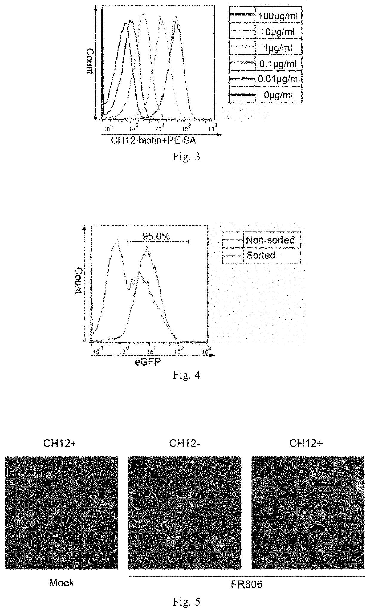
[0143]CH12-biotin was diluted to different concentrations (100 μg / ml, 10 μg / ml, 1 μg / ml, 0.1 μg / ml, 0.01 μg / ml, 0 μg / ml) in PBS containing 1% FBS, incubated with T cells expressing eGFP-F2A-FR806 for 45 min, and washed by PBS. The secondary antibody, PE-SA (ebioscience) was diluted at 1:300 in the medium, and resuspended cells were added and incubated for 45 min. Cells were washed twice with PBS and subjected to flow analysis. The results of flow analysis are showe...

example 3

R806-Positive T Cells with CH12-Biotin

[0144]1×10⁷ T cells expressing eGFP-F2A-FR806 were taken, washed with PBS, incubated with CH12-biotin (10 μg / ml, diluted with PBS containing 1% FBS) for 45 min at 4° C. and washed with PBS. Anti-Biotin sorting beads (purchased from Meitian Company) were added. T cells expressing FR806 were sorted according to the procedure provided with the sorting magnetic bead. Suitable amounts of the cells before and after sorting were taken and subjected to flow analysis. The results are shown in FIG. 4, demonstrating that, after binding to CH12-biotin, the T cells expressing FR806 can be effectively sorted by anti-Biotin sorting magnetic beads, and the positive rate of sorting is up to 95%.


PUM

PropertyMeasurementUnit
concentrationaaaaaaaaaa
volumeaaaaaaaaaa

Abstract

Provided are a fusion protein comprising an antibody binding area and an endocytic functional area, the encoding nucleic acid of the protein, an expression vector of same, a host cell thereof, and an immune effector cell expressing the fusion protein or the endocytic functional area or further expressing a chimeric antigen receptors. Also provided are an immunoconjugate comprising a cell-killing part and an antibody conjugate in a specifically-binding immune effector cell or an antibody of the endocytic functional area, a reagent kit and uses of the immunoconjugate, and a method for specifically removing, selecting, or enriching and detecting the immune effector cell.

Description

TECHNICAL FIELD[0001]The invention relates to the field of immunotherapy. In particular, the present invention relates to a fusion protein for controlling chimeric antigen receptor immune effector cells or TCR-T cells and uses thereof.BACKGROUND[0002]In recent years, great progress has been achieved in adoptive cell therapy (ACT), such as CAR-T and TCR-T against malignant tumors, among which the development of CAR-T therapy is the most significant.[0003]However, with the development of clinical trials of CAR-T cell therapy, there are many serious side effects, such as cytokine storms, off-target effects, etc. When serious adverse reactions occur, if the CAR-T cells are not inhibited in time, serious adverse, even life-threatening reactions will be incurred. Therefore, when using CAR-T treatment, it is necessary to introduce a safety switch at the same time, so that, when life-threatening reactions are incurred after CAR-T cells are used in a patient, the CAR-T cells in the body can ...

Claims


Application Information

Patent Timeline
Patent Type & Authority Applications(United States)
IPC IPC(8): C12N15/62C07K14/725C07K14/705C07K14/71C07K14/82C07K16/28C07K16/30C07K16/32C12N15/85C12N5/0783G01N33/537G01N33/547G01N33/569G01N33/577
CPCC07K14/70532C07K14/70578C07K14/70596C07K14/82C07K16/2803C07K16/2818C07K16/2827C07K16/2863C07K16/2878C07K16/2887C07K16/303C07K16/32C12N5/0636G01N33/56966C07K2319/02A61K39/4611A61K2239/31A61K2239/38A61K39/464412A61K2239/48A61K39/4631A61P35/00G01N33/56972A61K47/6903A61K47/6949C07K14/705C07K14/7051C07K14/71C07K2317/34C07K2319/01C07K2317/622C07K2319/03C07K2319/33C12N2510/00A61K2039/572A61K2300/00C07K19/00C12N5/10C12N15/62C12N15/85G01N33/537G01N33/547G01N33/577C07K16/30
Inventor LI, ZONGHAIWU, XIUQIWANG, HUAMAOJIANG, HUASHI, BIZHI
Owner CRAGE MEDICAL CO LTD
Learn More