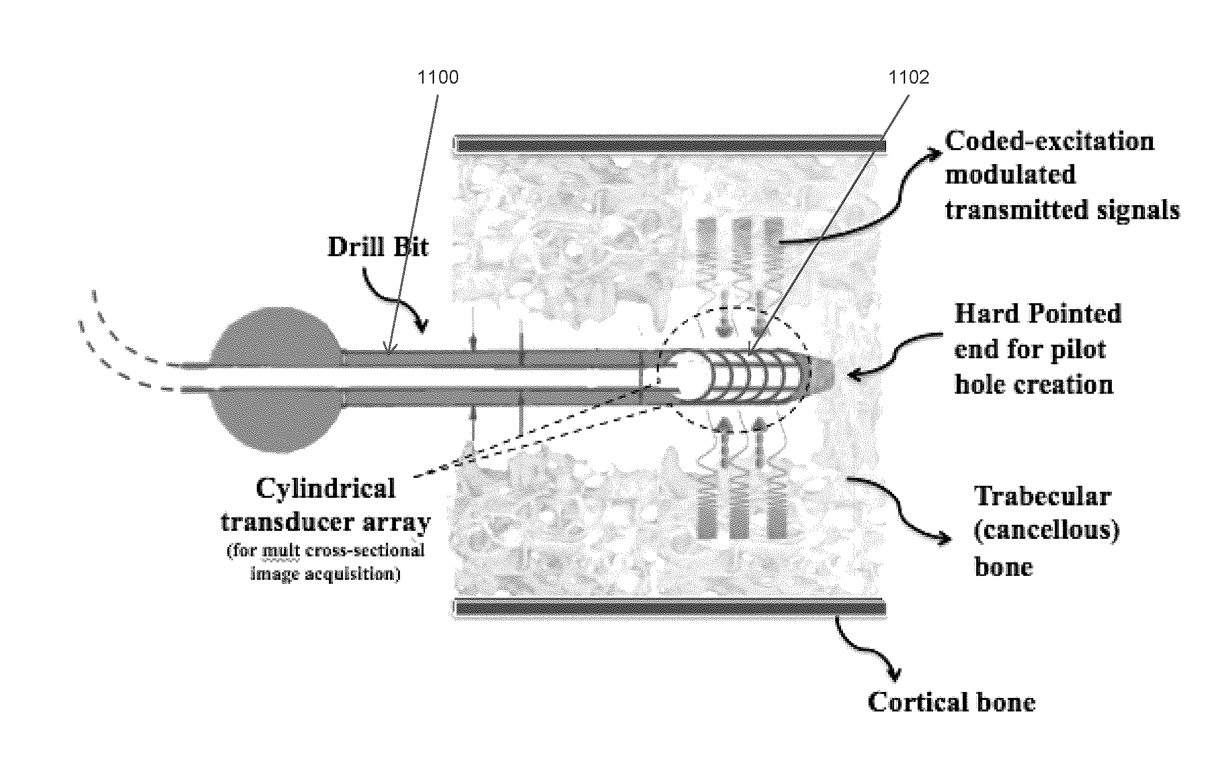
Ultrasonic signal processing for bone sonography
a technology of ultrasonic signal and bone sonography, which is applied in the field of ultrasonic signal processing and ultrasonic bone sonography, can solve the problems of difficult to decipher the image from the reflected ultrasound signal, difficult to obtain desired information by ultrasound imaging, and high frequency is susceptible to signal penetration depth loss, so as to achieve the effect of increasing the signal-to-noise ratio
- Summary
- Abstract
- Description
- Claims
- Application Information
AI Technical Summary
Problems solved by technology
Method used
Image

Examples
example 1
Comparing Methods of Coded-Excitation Signal Processing for Bone Sonography
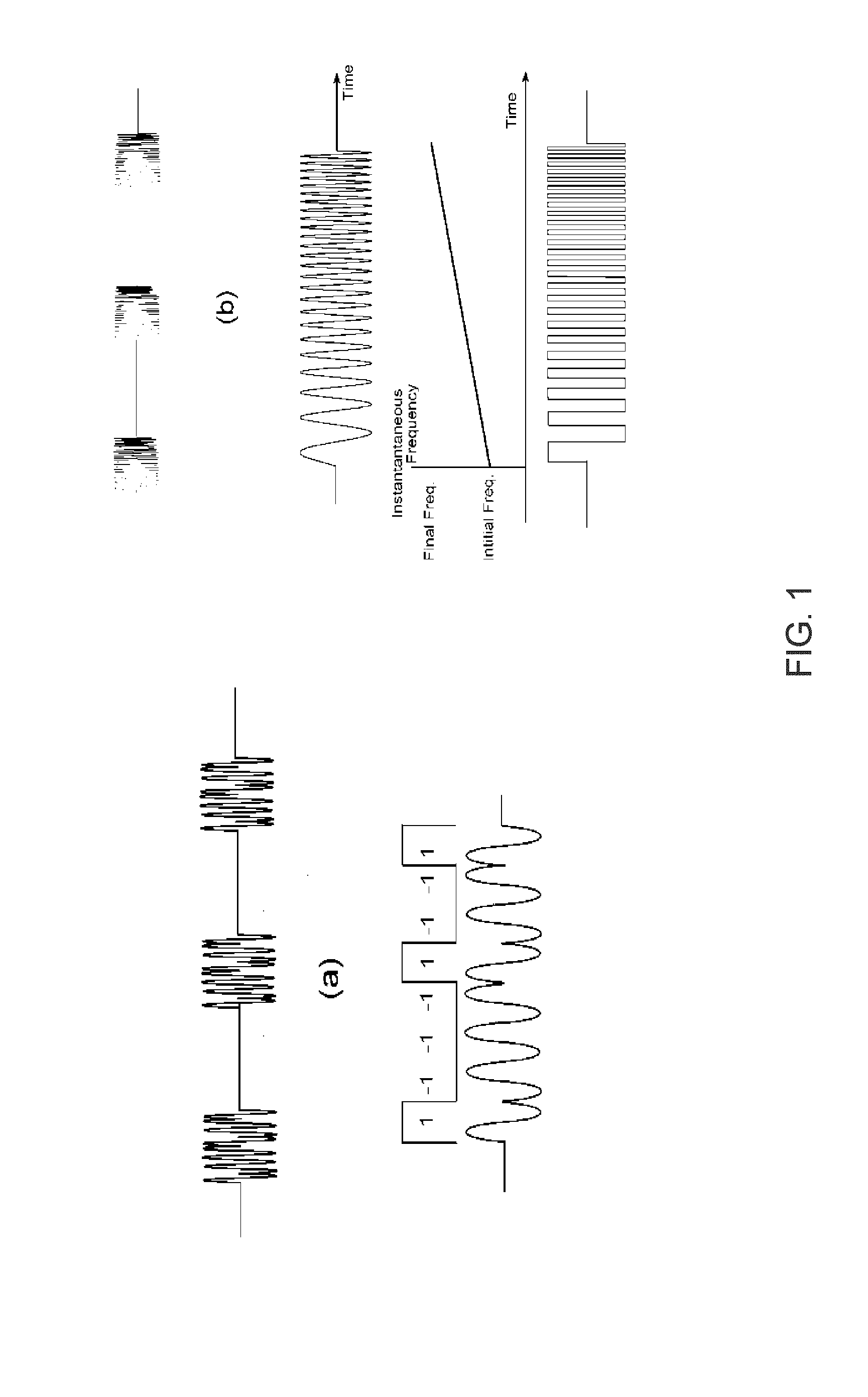
[0110]In Example 1 coded excitation methods, in particular Chirp modulation and Golay codes (GC), were found to enhance quality of ultrasound images by increasing ultrasonic signal-to-noise ratios (SNR) while preserving resolution.
[0111]Three signal compression techniques were compared to one another by directing ultrasound pulses toward two substrates: i) a 1.5 cm thick human cancellous bone placed on top of a glass microscopic glass (FIG. 17A) and ii) a 1.5 cm thick human cancellous bone placed on top of a cortical bone layer (FIG. 17B). Pulse-echo experiments were performed on each substrate to determine whether detectable signals could be observed and distinguished from the background signal received from the cancellous bone. Experimental design is illustrated in FIG. 17C.
[0112]Referring to FIG. 18A-C, three signal compression techniques were compared: i) single sinusoidal pulse, ii) Chirp modulation, and...
example 2
Enhancement of Signal to Noise Ratio in Modulated Ultrasound Signals
[0113]In Example 2, three SNR (Signal-to-Noise Ratio) enhancement methods were compared in simulation models based on using a 2 MHz transducer with 80% bandwidth.
[0114]Referring to FIG. 19, the response of one cycle sine pulse (2MHz) after 20 times was averaged. The effect of averaging enhanced the SNR by square root of number of averages: √{square root over ( N; where N is the number of averaging. Square root function reaches a plateau relatively quickly, thus experimental work was required to determine a reasonable averaging outcome, without spending too much time taking more and more signals. Experimentation suggested that N=20-50 was a reasonable number.
[0115]Referring to FIG. 20, the effect of chirp modulation employing a time duration of 0.25 ms (milliseconds) with a frequency sweeping from 0.5 to 3.5 MHz (Bandwidth of 3 MHz) was considered. Theoretically, the SNR enhancement was proportional to square root of...
PUM
 Abstract
Description
Claims
Application Information
 - R&D
- Intellectual Property
- Life Sciences
- Materials
- Tech Scout
- Unparalleled Data Quality
- Higher Quality Content
- 60% Fewer Hallucinations
Browse by: Latest US Patents, China's latest patents, Technical Efficacy Thesaurus, Application Domain, Technology Topic, Popular Technical Reports.
© 2025 PatSnap. All rights reserved.Legal|Privacy policy|Modern Slavery Act Transparency Statement|Sitemap|About US| Contact US: help@patsnap.com