
Establishment method and applications of microfluidic-chip-based tumor infiltration model

A microfluidic chip and chip technology, applied in the field of cell biology research, can solve the problem that the movement characteristics of circulating tumor cells are in a blank stage

Inactive Publication Date: 2018-06-05
DALIAN INST OF CHEM PHYSICS CHINESE ACAD OF SCI
View PDF2 Cites 2 Cited by
  • Summary
  • Abstract
  • Description
  • Claims
  • Application Information

AI Technical Summary

Problems solved by technology

At present, the simulation of tissue engineering using microfluidic chips, especially the simulation of brain microvascular inflammation and the research and analysis of the movement characteristics of circulating tumor cells in the inflammatory site are still in a blank stage.

Method used


Image

  • Establishment method and applications of microfluidic-chip-based tumor infiltration model
  • Establishment method and applications of microfluidic-chip-based tumor infiltration model
  • Establishment method and applications of microfluidic-chip-based tumor infiltration model

Examples


Embodiment 1

[0041] Establishment of Vascular Inflammation Lesion Model Using Brain Microvascular Endothelial Cells

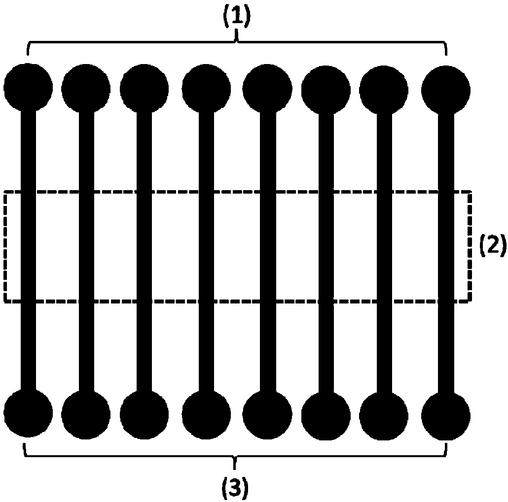
[0042] Using the microfluidic chip designed and manufactured by the laboratory, the structure is as follows: figure 1 shown. Prepare a type I rat tail collagen working solution with a concentration of 0.1mg / mL, inject it into the chip through the cell inlet, and let it stand overnight at room temperature. After 24 hours, remove the collagen working solution, and wash the channel 3 times with the cell culture solution. Dilute the brain microvascular endothelial cells to a concentration of 5×10 6 Cells / mL cell suspension, add 100 μL of cell suspension into the chip through the cell inlet, under the modification of collagen, the cells quickly adhere to the wall and spread evenly on the bottom of the cell culture chamber, when the cells are observed under the optical microscope When the cells are evenly distributed in the culture room, immediately move the chip into the carbo...

Embodiment 2

[0044] Characterization of Intracellular Functional Proteins in a Cerebral Microvascular Inflammation Model

[0045] Using the microfluidic chip designed and manufactured by the laboratory, the structure is as follows: figure 1 shown. After chip modification, the same cell inoculation and culture methods as in Example 1 were used to establish a cerebral microvascular inflammation model. After 24 hours of stimulation, immunofluorescence staining was performed on the cells, and the monitored proteins were carbon monoxide synthase (eNOS), von Willebrand factor (vWF), integrin β4 (Intβ4), vinculin, and endothelial cell adhesion molecule- 1 (VCAM-1) and intercellular adhesion molecule-1 (ICAM-1), photographed under a fluorescent microscope, and recorded the expression of the corresponding proteins, the results are as follows Figure 4 shown.

Embodiment 3

[0047] Adhesion and rolling of lung cancer cells on the surface of brain microvascular endothelial cells under fluid shear force

[0048] Using the microfluidic chip designed and manufactured by the laboratory, the structure is as follows: figure 1 shown. After chip modification, the same cell inoculation and culture methods as in Example 1 were used to establish a cerebral microvascular inflammation model. Then the lung cancer cells were digested and diluted into a cell suspension with a concentration of 1×10 6 cells / mL, through the cell entrance into the channel, the microscope continuously photographed the adhesion and rolling phenomenon of lung cancer cells on the surface of brain microvascular endothelial cells, and counted the number of lung cancer cells that adhered and rolled. The results are as follows: Figure 5 shown. Figure 6 Adhesion and rolling of lung cancer cells on the surface of brain microvascular endothelial cells under the action of different molecules...


PUM


Abstract

The invention discloses an establishment method and applications of a microfluidic-chip-based tumor infiltration model, wherein the microfluidic chip comprises a cell inlet, a cell culture chamber anda waste liquid outlet, the cell inlet is connected to the upper portion of the cell culture chamber, and the waste liquid outlet is connected to the lower portion of the cell culture chamber. The model establishment steps comprise: (1) chip modification, (2) inoculation and culture of cells in the chip, and (3) establishment of an inflammatory brain microvessel model. According to the present invention, the model is particularly used for the brain microvascular inflammation part, and can simulate the adhesion and the rolling of tumor cells on the surface of brain microvascular endothelial cells under the blood flowing condition; and with the method, the simulation of the brain microvascular lesion under the inflammatory condition and the enrichment of the circulating tumor cells at the lesion part under the blood flowing condition can be integrated so as to simulate the infiltration of tumor cells in blood vessels.

Description

technical field [0001] The invention relates to the technical field of applying microfluidic chip technology to cell biology research of tissue bionics and real-time monitoring, in particular to a method and application of a tumor infiltration model based on a microfluidic chip. Background technique [0002] Tumor brain metastasis is a complex physiological process. The distant metastasis of tumor cells from the lesion site includes multiple steps, such as detachment of tumor cells from tumor tissue, in situ growth and proliferation, invasion and extravasation, passing through the blood vessel wall into the circulatory system, Adhesion to the blood vessel wall, infiltration into another tissue or organ at the far end, each step is inseparable from the regulation and support of the tumor microenvironment. Among the fatal cases of tumor-related diseases, the mortality rate of metastatic tumors is as high as 90%, and clinical statistics show that the metastasis rate and mortali...

Claims


Application Information

Patent Timeline
Patent Type & Authority Applications(China)
IPC IPC(8): C12M3/00C12N5/071C12Q1/06
CPCC12M23/16C12N5/0691C12N2501/25G01N33/5029G01N33/5064
Inventor 秦建华许慧李中玉
Owner DALIAN INST OF CHEM PHYSICS CHINESE ACAD OF SCI
Learn More