A three-dimensional cell capture and release chip and its preparation method
A three-dimensional structure and cell technology, applied in chemical instruments and methods, laboratory containers, laboratory utensils, etc. and other problems, to achieve the effect of high efficiency, short capture time and simple processing technology
- Summary
- Abstract
- Description
- Claims
- Application Information
AI Technical Summary
Problems solved by technology
Method used
Image
Examples
Embodiment 1
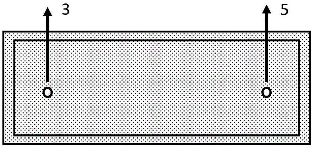
[0037] Such as Figure 1-2 As shown, a three-dimensional cell capture and release chip includes an upper cover 1 and a lower substrate 2, the back of the upper cover 1 is etched with a microfluidic channel 4, and the microfluidic channel 4 of the cover 1 is two An inlet 3 and an outlet 4 are respectively arranged at the ends, a layer of chitosan film 6 is provided on the substrate 2, and the cover sheet 1 and the substrate 2 are bonded by an adhesive.
[0038] The preparation method of the cell capture and release chip with the above three-dimensional structure, the steps are as follows:
[0039] (a) Select glass, silicon wafer or quartz as the material of the cover and substrate, and clean the selected cover and substrate to remove impurities such as oil on the surface;
[0040] (b) Dry the cleaned cover and substrate in an oven at 50°C to 100°C. After drying, adhere a chemical-resistant mask to the upper and lower surfaces of the cover, and cut it by laser. Cut out part of...
Embodiment 2
[0047] A method for preparing a three-dimensional cell capture and release chip, the steps are as follows:
[0048] (a) Select the material or glass of the cover and substrate as glass, silicon wafer or quartz, and clean the selected cover and substrate to remove impurities such as oil stains on the surface;
[0049] (b) Dry the cleaned cover and substrate in an oven at 50°C to 100°C. After drying, adhere a chemical-resistant mask to the upper and lower surfaces of the cover, and cut it by laser. Cut out part of the area to be etched, remove the anti-corrosion mask cut out, and obtain the area to be etched;
[0050] (c) Put the processed cover sheet into the etching solution vertically and slowly at the rate of 0.01mm / min or 0.1mm / min or 0.25mm / min or 0.75mm / min or 1mm / min through the fixture for etching Etching, etch for 1 hour, because there is a sequence of entering the etching solution, so the etching time is different, resulting in different etching depths, so the microflu...
Embodiment 3
[0056] A method for capturing and releasing cells based on a chip with a three-dimensional structure, the steps of which are as follows:
[0057] (1) if Figure 3-4 As shown, the capture of cells:
[0058] (a) inject the liquid sample containing ctc circulating tumor cells to be detected into the chip of Example 1 through the syringe pump at a rate of 0.51ul / min from the inlet hole;
[0059] (b) Continue to inject phosphate buffer solution (i.e. PBS solution) at the inlet, and inject at a speed of 50ul / min for cleaning;
[0060] (c) Inject tracer substances (such as: fluorescent dyes such as DAPI or Hoechst dyes that stain the nucleus) at the entrance to identify target cells;
[0061] (d) Inject phosphate buffer solution at the inlet again for cleaning, and place the chip under a microscope to observe the captured target cells;
[0062] (2) if Figure 5-6 As shown, the cells release:
[0063] (a) The chip captures cells with a specific capture size stuck in the correspon...
PUM
 Abstract
Description
Claims
Application Information
 - R&D Engineer
- R&D Manager
- IP Professional
- Industry Leading Data Capabilities
- Powerful AI technology
- Patent DNA Extraction
Browse by: Latest US Patents, China's latest patents, Technical Efficacy Thesaurus, Application Domain, Technology Topic, Popular Technical Reports.
© 2024 PatSnap. All rights reserved.Legal|Privacy policy|Modern Slavery Act Transparency Statement|Sitemap|About US| Contact US: help@patsnap.com


