
Equipment and method for dental X-ray tomography

A tomography and X-ray technology, applied in the field of radiation imaging, can solve the problems of high economic burden, high cost and large radiation dose in dental clinics and patients, and achieve the effect of high market application potential.

Active Publication Date: 2013-01-09
TSINGHUA UNIV
View PDF6 Cites 12 Cited by
  • Summary
  • Abstract
  • Description
  • Claims
  • Application Information

AI Technical Summary

Problems solved by technology

However, due to the use of large-area flat-panel detectors in CBCT equipment, its cost is relatively high, and the economic burden is relatively large for dental clinics and patients; and the radiation dose of CBCT is relatively large, which has a great impact on controlling the average dose level of the public. Negative impact; Finally, due to the limited spatial resolution of the flat panel detector used in CBCT, the imaging accuracy of CBCT is much lower than that of X-ray CR or DR imaging

Method used


Image

  • Equipment and method for dental X-ray tomography
  • Equipment and method for dental X-ray tomography
  • Equipment and method for dental X-ray tomography

Examples


Embodiment Construction

[0029] Embodiments of the present invention are described in detail below, examples of which are shown in the drawings, wherein the same or similar reference numerals designate the same or similar elements or elements having the same or similar functions throughout. The embodiments described below by referring to the figures are exemplary and are intended to explain the present invention and should not be construed as limiting the present invention.

[0030] In describing the present invention, it should be understood that the terms "center", "longitudinal", "transverse", "length", "width", "thickness", "upper", "lower", "front", " Orientation or position indicated by "back", "left", "right", "vertical", "horizontal", "top", "bottom", "inner", "outer", "clockwise", "counterclockwise", etc. The relationship is based on the orientation or positional relationship shown in the drawings, and is only for the convenience of describing the present invention and simplifying the descrip...


PUM


Abstract

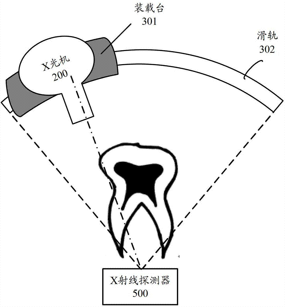
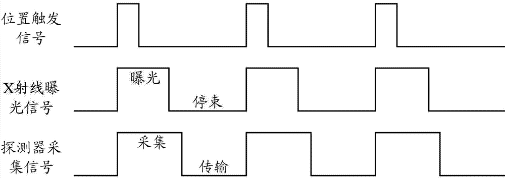
The invention provides equipment and a method for dental X-ray tomography, wherein the equipment comprises a scanning control module, an X-ray machine, a mechanical scanner, an image acquisition control module, an X-ray detector and a tomographic image reconstruction module as well as an image display module, wherein the scanning control module is used for emitting an exposure pulse signal to control the exposure of the X-ray machine and emitting an acquisition pulse signal to control the acquisition work of the image acquisition control module; the mechanical scanner is used for driving the X-ray machine to move to different positions to finish the tomographic scanning process; the image acquisition control module is used for converting, digitalizing and transmitting detection results of the X-ray detector; the tomographic image reconstruction module is used for reconstructing an acquired multi-angle X-ray perspective image to obtain a multi-position cross-sectional image of a tissue to be imaged; the position to be imaged is arranged between the X-ray machine and the X-ray detector, and X rays emitted by the X-ray machine always pass through the X-ray detector. The equipment for dental X-ray tomography, provided by the invention, has the advantages of low cost, high imaging speed and good imaging effect.

Description

technical field [0001] The invention relates to the field of radiation imaging, in particular to a dental X-ray tomographic imaging device and method. Background technique [0002] X-ray imaging technology has become an indispensable imaging technology in the diagnosis of dental oral diseases. Relying on X-ray imaging technologies such as X-ray perspective imaging, panoramic (Panoramic), cone-beam CT (Cone-beam CT, CBCT) and other X-ray imaging technologies have gradually met various Dental diagnostic needs for oral diseases. There are three different technical means of X-ray fluoroscopic imaging: film imaging, digital X-ray imaging technology (Computed Radiography, CR) and direct digital X-ray imaging technology (Digital Radiography, DR). However, due to the advantages of simple technology, easy operation, cheap equipment, and low dose, it is widely used in intraoral dental X-ray imaging at present. In order to be able to complete the imaging of all the teeth and maxilla ...

Claims


Application Information

Patent Timeline
IPC IPC(8): A61B6/14A61B6/03
Inventor 李亮陈志强赵自然唐志宏张丽
Owner TSINGHUA UNIV
Learn More